
Fig. 2.
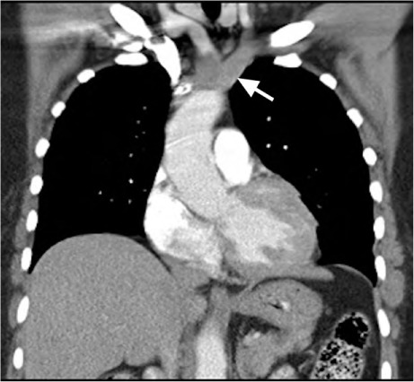
Computed tomogram (coronal view) with intravenous contrast medium shows a large aortic intimal sarcoma that was initially mistaken for thrombus. The tumor involves most of the aortic arch, extending superiorly into the proximal portions of the brachiocephalic trunk and left subclavian artery (arrow) and into the proximal descending aorta.
Supplemental motion image (4MB, m1v) is available for Fig. 2.