Fig. 3.
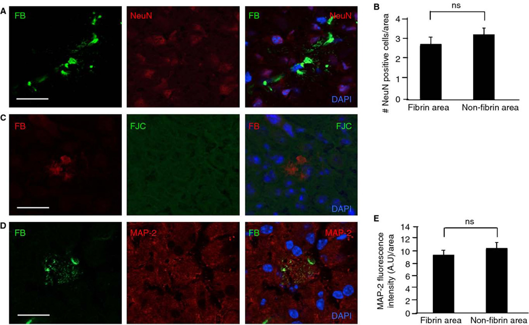
Fibrin deposits in plg−/− mouse brains do not significantly affect neuronal health. Coronal brain tissue sections from plg−/− animals (n=12 mice, 3–5 sections per animal) were stained, and neuronal markers in cortical areas were quantified. A) Co-immunostaining for fibrin (FB, green) and neuronal nuclei (NeuN, red) does not show a significant loss of neurons in proximity to fibrin deposits. B) There is no significant difference in number of NeuN-positive cells in fibrin vs. non-fibrin immunoreactive areas. C) Fibrin deposition does not correlate with neuronal cell death as visualized by co-immunostaining for fibrin (red) and Fluoro-Jade C (FJC, green). D) Co-immunostaining for fibrin (green) and microtubule-associated protein-2 (MAP-2, red) indicates that fibrin does not have a significant effect on dendritic density. E) There is no significant difference in MAP-2 fluorescence intensity in fibrin vs. non-fibrin immunoreactive areas. Differences in neuronal markers (NeuN and MAP-2) between fibrin and non-fibrin immunoreactive areas were evaluated by Student’s t-test. Values are presented as mean ± SEM. Scale bars=20 µm. DAPI, blue.

