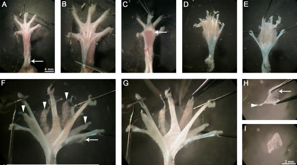
Figure 1. Hind-limb lumbrical muscles can be easily dissected.
Immediately after culling, the hind-limbs are removed and overlying skin detached. The leg (left in this figure) is then pinned out ventral side up (A), and the remaining plantar skin dissected away revealing the tendons in the foot. The overlying FDB tendon is cut transversely close to the ankle, lifted up towards the digits, and removed completely by cutting the distal ends of the tendon at the level of the metatarsophalangeal joints (A, arrow). The underlying FDL tendon, from which the lumbricals originate, is now visible (B). This tendon is also cut proximally and pulled back towards the digits (C). The tendons, along with connective tissue and the distal ends of the lumbricals, are cut close to the base of the digits (C, arrow) and the plantar muscle mass lifted out. The FDL tendon is then spread and tautly pinned ventral side up (E, not D). The connective tissue is carefully cleared, revealing the underlying lumbricals (arrowheads) between the tendon ends (from medial to lateral, the first to fourth) (F). The FDL muscles (F, arrow) can be detached to increase visibility of the lumbricals (G). Excess connective tissue is removed by blunt dissection in the regions between the lumbricals and the tendons, before fixation. Lumbrical muscles are excised and further connective tissue (H, arrow) removed. Once cleaned up, the distal FDB tendon (arrowhead) is disconnected, and the muscle is ready for staining (I). Lumbrical muscles were dissected from a two month-old wild-type mouse in this figure.

