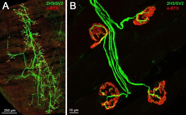
Figure 2. The entire innervation pattern of lumbrical muscles can be imaged and NMJs examined.
A. A representative confocal micrograph of a wild-type mouse hind-limb lumbrical muscle at post-natal day 15. The entire innervation pattern is visible in the thin, whole-mount preparation, obviating the need for muscle sectioning. Two separate “collapsed” images were tiled together to produce this figure. B. NMJs can be viewed in fine detail, allowing accurate characterisation of a number of phenotypes (see Fig. 3). This image shows example NMJs from a two month-old wild-type mouse. For this and the subsequent figure, nervous tissue is stained green (2H3, neurofilament, and SV2, synaptic vesicle 2) and post-synaptic AChRs are stained red (-BTX, -bungarotoxin). 10× and 63× objectives were used to produce the images in panels A and B, respectively.

