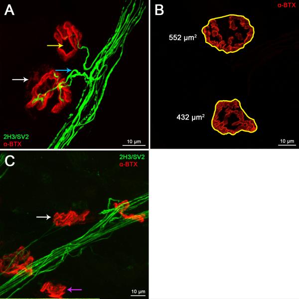
Figure 3. Lumbrical NMJs can be accurately assayed for developmental and denervation phenotypes.
A. In early post-natal development, NMJs undergo a number of maturational processes that can be assessed using confocal microscopy. Synapse elimination can be scored by calculating the percentage of NMJs that remain polyinnervated (A, blue arrow), and the plaque-to-pretzel transition can be measured up to one month post-birth by counting the number of perforations per post-synaptic endplate (A, yellow arrow). These maturational processes occur during the first few post-natal weeks in wild-type animals, and can be studied in neurological disease models to determine defective neuromuscular development. B–C. Synaptic growth and neurodegeneration can also be evaluated in wild-type and disease model mice. Confocal Z-stack images can be used to measure post-synaptic area and average fluorescence intensity (B), while loss of lower motor neuron connectivity can be determined by scoring the percentage of NMJs displaying a lack of overlap in pre-and post-synaptic staining (C). The white arrow in panel A identifies a partially denervated synapse, while the purple and white arrows in panel C highlight fully and partially denervated NMJs, respectively. Panels A and C show NMJs from a two month-old mouse model of the peripheral neuropathy Charcot-Marie-Tooth disease type 2D (GarsNmf249/+), and panel B depicts two month-old wild-type endplates. A 63x objective was used to produce the images for this figure.

