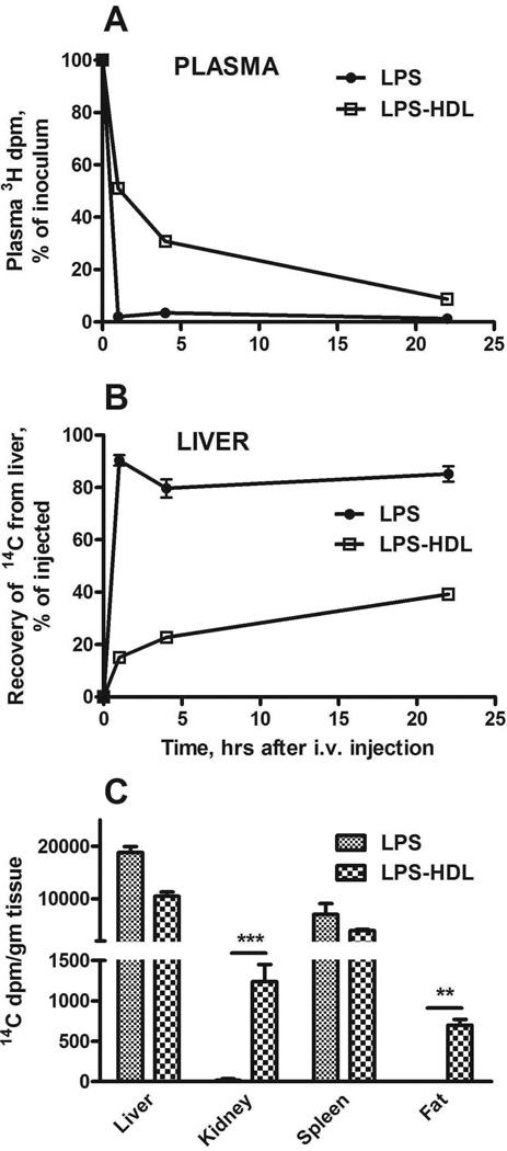
Figure 1. Plasma clearance and tissue uptake of radiolabeled LPS and HDL-LPS.
LPS or HDL-LPS, each containing 5 µg [3H/14C]LPS, was injected via the lateral tail vein. A and B show the time course for disappearance of LPS and LPS-HDL from the plasma (A) and their uptake by the liver (B). C compares the tissue distribution of LPS and LPS-HDL 22 hrs after i.v. injection. n = 3–6 mice/timepoint. Error bars = 1 SEM. Data combined from 3 experiments with similar results. ** p <0.01, *** p < 0.001.

