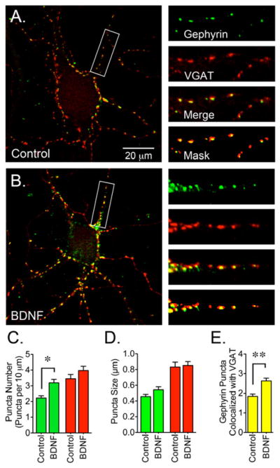
Figure 2. BDNF-Dependent Increase in Gephyrin Clustering.

(A) Control or (B) BDNF-treated cultures were co-stained with gephyrin (3B11 antibody, detecting the E-domain, green) and VGAT (red) antibodies. (C) BDNF-treated cultures (50 ng/ml for 2 days starting at 8 DIV) showed an increase in the number of gephyrin clusters. (D) Gephyrin and VGAT puncta size. (E) The number of gephyrin clusters colocalizing with VGAT augmented after BDNF treatment. Data are presented as the mean ± S.E.M. of 15 cells from 3 independent cultures. Green bars represent gephyrin, red bars represent VGAT and yellow bars represent gephyrin clusters that colocalize with VGAT (per 10 μm). BDNF triggers a significant increase in the number and synaptic localization of gephyrin clusters (*p<0.05 or **p<0.01 compared to control cultures by Student’s t-test). Scale bar in A represents 20 μm and the areas within the white boxes are shown at a higher magnification and represent 30 μm.
