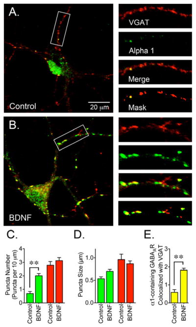
Figure 6. BDNF Promotes the Colocalization of GABAAR and VGAT.

(A) Control or (B) BDNF-treated cells were co-stained with a VGAT antibody (red) in combination with an α1 subunit antibody (green). (C) BDNF treatment (50 ng/ml for 2 days) resulted in a significant increase in the number of GABAAR clusters. (D) Cluster size for α1-containing GABAAR and VGAT. (E) The number of α1 clusters that colocalized with VGAT was increased after BDNF treatment. Data presented are the mean ± S.E.M. of 15 cells from 3 independent cultures. Green bars represent values for the α1 subunit, red bars represent VGAT values and yellow bars represent GABAAR clusters that colocalize with VGAT (per 10 μm). BDNF triggers a significant increase in the number of α1 clusters and also increases the colocalization of GABAAR and VGAT (**p<0.01 compared to control cultures by Student’s t-test). Scale bar represents 20 μm and the areas within the white boxes are shown at a higher magnification and represent 30 μm.
