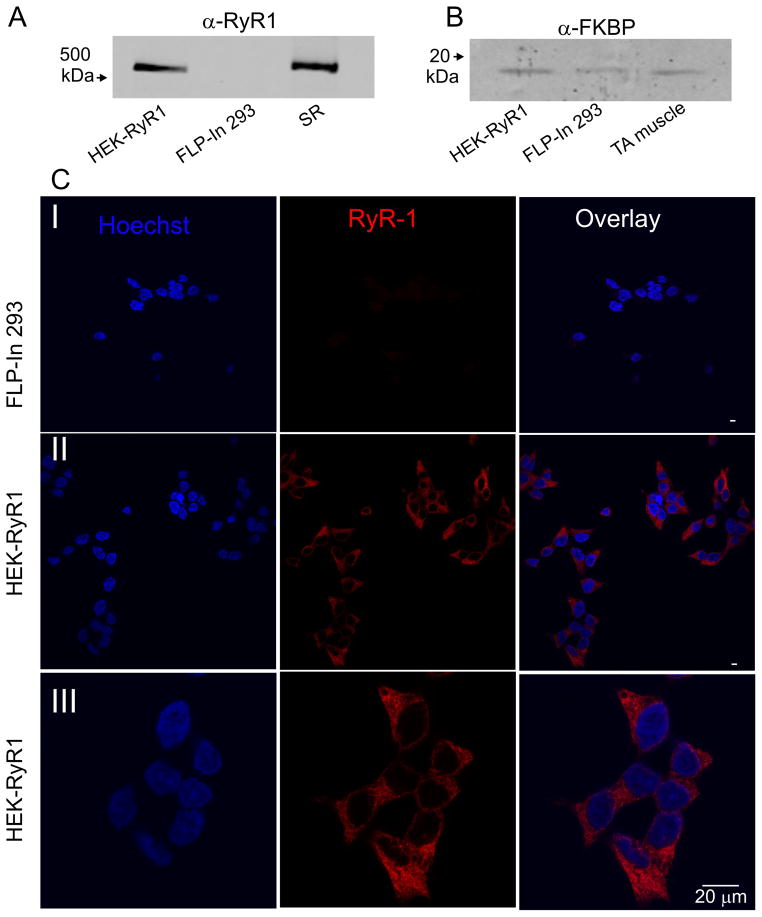
Figure 1. RyR1 and FKBP expression in RyR1 stable Flp-In cells.
A) Representative western blot analysis of 10 μg each of HEK-RyR1 and control Flp-In 293 cell lysates probed with anti-RyR antibody. A sarcoplasmic reticulum (SR) preparation (5 μg) from mouse skeletal muscle was used as a positive control. B) Representative western blot analysis of 10 μg each of HEK-RyR1 and control Flp-In 293 cell lysates probed with anti-FKBP (1:100) antibody. A tibialis anterior (TA) mouse muscle homogenate preparation (10 μg) was used as a positive control for FKBP12. C) Control Flp-In 293 cells (Panel I) and HEK-RyR1 cells (Panels II and III) showing Hoechst 34580 nuclear staining alone (left), RyR1 immunostaining alone (middle) and overlay (right).

