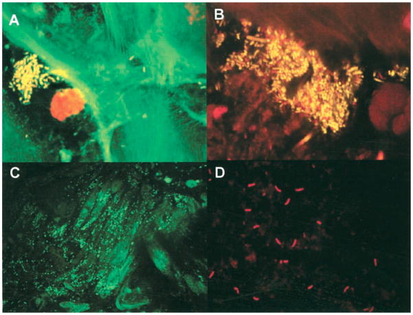
Figure 1.
In situ visualization of bacteria in esophageal mucosal biopsies. A and B: Confocal sections of mucosal biopsy specimens stained for cell viability, containing mixtures of living (yellow) and dead (red) organisms. Microcolony and aggregate formation can be seen in mucosal samples from control subjects (A) and from patients with Barrett’s esophagus (B). C and D: Fluorescence light micrographs of transverse sections of Barrett’s esophageal mucosae showing colonization by streptococci (C) and Campylobacter species (D), using 16S ribosomal RNA oligonucleotide probes labeled with FITC and cy3. Figure originally published in Journal of Clinical Infectious Diseases15 and permission to reuse of the figure is granted by the Journal.

