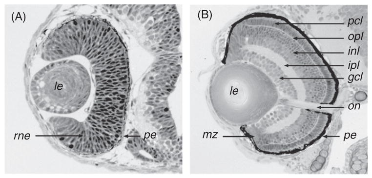
Fig 2.

Histology of the zebrafish retina. (A) A section through the zebrafish eye during early stages of neurogenesis at approximately 36 hpf. At this stage, the retina mostly consists of two epithelial layers: the pigmented epithelium and the retinal neuroepithelium. Although some retinal cells are already postmitotic at this stage, they are not numerous enough to form a distinct layer. (B) A section through the zebrafish eye at 72 hpf. With the exception of the marginal zone, where cell proliferation will continue throughout the lifetime of the animal, retinal neurogenesis is mostly completed. The major nuclear and plexiform layers, as well as the optic nerve and the pigmented epithelium, are well differentiated. gcl: ganglion cell layer; inl: inner nuclear layer; ipl: inner plexiform layer; le: lens; mz: marginal zone; on: optic nerve; opl: outer plexiform layer; pcl: photoreceptor cell layer; pe: pigmented epithelium; rne: retinal neuroepithelium.
