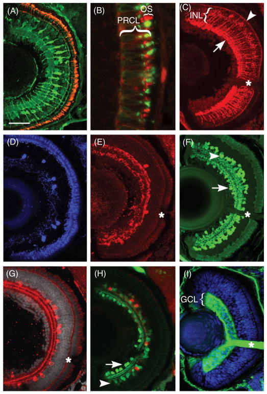
Fig 3.
Transverse sections through the center of the zebrafish eye reveal several major retinal cell classes and their subpopulations. (A) Anti-rod opsin antibody detects rod photoreceptor outer segments (red), which are fairly uniformly distributed throughout the outer perimeter of the retina by 5dpf. On the same section, an antibody to carbonic anhydrase labels cell bodies of Müller glia in the INL as well as their radially oriented processes. (B) A higher magnification of the photoreceptor cell layer shows the distribution of rod opsin (red signal) and UV opsin (green signal) in the outer segments (OSs) of rods and short single cones, respectively. (C) A subpopulation of bipolar cells is detected using antibody directed to protein kinase C-β (PKC). While cell bodies of PKC-positive bipolar neurons are situated in the central region of the INL, their processes travel radially into the inner (arrow) and outer (arrowhead) plexiform layers, where they make synaptic connections. (D) Tyrosine hydroxylase-positive interplexiform cells are relatively sparse in the larval retina. (E) Similarly, the distribution of neuropeptide Y is limited to only a few cells per section. (F) The distribution of GABA, a major inhibitory neurotransmitter. GABA is largely found in amacrine neurons in the INL (arrowhead), although some GABA-positive cells are also found in the GCL (arrow). (G) Choline acetyltransferase, an enzyme of acetylcholine biosynthetic pathway, is restricted to a relatively small amacrine cell subpopulation. (H) Antibodies directed to a calcium-binding protein, parvalbumin, recognize another fairly large subpopulation of amacrine cells in the INL (green, arrowhead). Some parvalbumin−positive cells localize also to the GCL and most likely represent displaced amacrine neurons (arrow). By contrast, serotonin-positive neurons (red) are exclusively found in the INL. (I) Ganglion cells stain with the Zn-8 antibody directed to neurolin, a cell surface antigen (Fashena and Westerfield, 1999). In addition to neuronal somata, strong Zn-8 staining exists in the optic nerve (asterisk). In all panels lens is left, dorsal is up. A–H show the retina at 5dpf, while I shows a 3dpf retina. Asterisks indicate the optic nerve. Scale bar equals 50 μm in A and C–I and 10 μm in B. dpf, days post fertilization; GCL, ganglion cell layer; INL, inner nuclear layer; OS, outer segments; PRCL, photoreceptor cell layer. Panels D, G, and H are reprinted from Pujic and Malicki (2004) with permission from Elsevier. (See Plate no. 8 in the Color Plate Section.)

