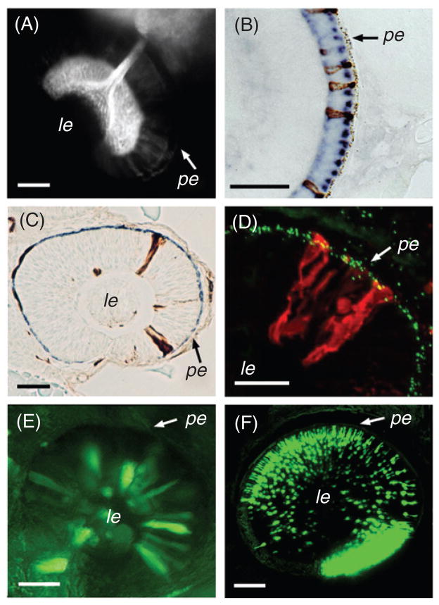
Fig 4.
Selected techniques available to study neurogenesis in the zebrafish retina. (A) DiI incorporation into the optic tectum retrogradely labels the optic nerve and ganglion cell somata. (B) A transverse plastic section through the zebrafish retina at 3 dpf. In situ mRNA hybridization using two probes, each targeted to a different opsin transcript and detected using a different enzymatic reaction, visualizes two types of photoreceptor cells. (C) A plastic section through a genetically mosaic retina at ca. 30 hpf. Biotinylated dextran-labeled donor-derived cells incorporate into retinal neuroepithelial sheet of a host embryo and can be detected using HRP staining (brown precipitate). (D) A transverse cryosection through a genetically mosaic zebrafish eye at 36 hpf. In this case, donor-derived clones of neuroepithelial cells are detected with fluorophore-conjugated avidin (red). The apical surface of the neuroepithelial sheet is visualized with anti-γ-tubulin antibody, which stains centrosomes (green). (E) GPF expression in the eye of a zebrafish embryo following injection of a DNA construct containing the GFP gene under the control of a heat-shock promoter. The transgene is expressed in only a small subpopulation of cells. (F) A confocal z-series through the eye of a living transgenic zebrafish, carrying a GFP transgene under the control of a rod opsin promoter (Fadool, 2003). Bright expression is present in rod photoreceptor cells (ca. 3 dpf). Scale bar, 50 μm. pe, pigmented epithelium; le, lens. Panel E reprinted from Malicki et al. (2002) with permission from Elsevier.

