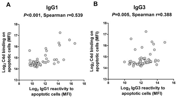
Figure 6. Correlation between complement activation and IgG subclasses reactivity to apoptotic cells.

The deposition of C4d on the surface of apoptotic Jurkat cells was detected by flow cytometry after complement activation in vitro with IgG purified from the 50 most reactive pre-transplant serum samples. C4d deposition is reported together with IgG1 (A) or IgG3 (B) reactivity to apoptotic cells measured in the same samples.
