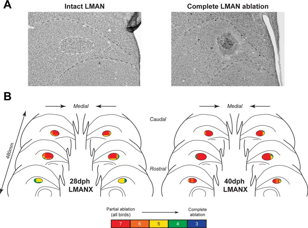
Figure 3. Overlay of bilateral coronal sections in both LMAN-ablation groups.
(A) Representative images of intact LMAN (outlined in white, from a CTL bird) and a complete LMAN ablation are shown (from a 28dph bird). (B) Bilateral coronal sections for both 28- and 40- dph LMANX groups show overlapping LMAN ablation sites in all LMANX birds (N=7 in both 28- and 40-dph groups). The position and amount of LMAN ablated for each bird were normalized relative to tracings of LMAN in CTL birds, and then combined to create the overlays. Color fills indicate regions of overlapping ablation across birds (e.g., all seven birds in each group received ablation in red-shaded regions of LMAN). Three birds in each group received complete LMAN ablation. The pattern of overlap indicates that LMAN ablation occurred most consistently in caudal and central LMAN.

