Figure 2.

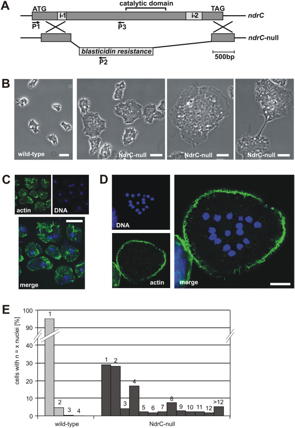
NdrC-null cells are impaired in cell division. A. Gene replacement of ndrC (DDB0219984) by a blasticidin-S resistance cassette. P1-P2 and P1-P3 indicate the primer combinations that were used initially to identify gene knockouts by PCR. B. Live-cell microscopy of wild-type (left) and ndrC-null cells. ndrC-null cells are much larger than wild-type cells, and often divide by traction-mediated cytofission (as shown in the right image). C. Fixed wild-type cells stained with TRITC-phalloidin for actin and TO-PRO-3 for DNA. D. Fixed ndrC-null cell stained with TRITC-phalloidin for actin and TO-PRO-3 for DNA. E. Histogram showing the percentage of cells carrying the indicated numbers of nuclei in wild-type and ndrC-null mutants. Cells were grown in Petri dishes, fixed, stained, and for each strain the nuclei of >500 cells were counted. The counting was repeated in an independent experiment with almost identical results. Bars, 10 μm.
