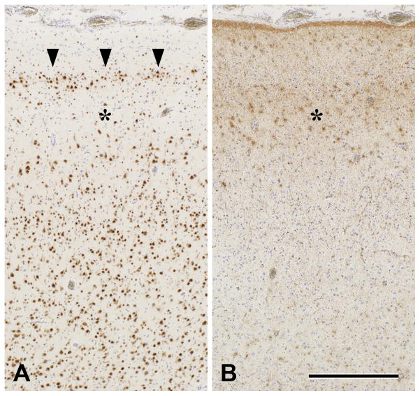
Fig. 8. Temporal lobe sclerosis observed in a 43-year-old man with mTLE since the age 24.
Pre-operative MRI showed T2/FLAIR high intensity change in the right atrophic hippocampus. FDG-PET revealed hypometabolism in the right temporal tip to mesial temporal structure. The patient underwent surgical intervention by combined amygdalohippocampectomy and anterior temporal lobectomy on the right side. Pathological examination of the resection specimen revealed hippocampal sclerosis (HS) with granule cell loss in the dentate gyrus and amygdala sclerosis as well as a feature of temporal lobe sclerosis (TLS) in the temporal neocortex shown in panels A and B. Based on the presence of TLS in association with HS, cortical abnormality in this case is consistent with one variant of FCD type IIIa. The patient is seizure free for more than 2 years after the operation (Engel class I).
A: An abnormal band of small and clustered “granular” neurons in the outer part of cortical layer 2 (arrowheads) and underlying paucicellular area (asterisk). NeuN immunohistochemistry counterstained with hematoxylin.
B: Laminar gliosis in cortical layers 2 and 3 (asterisk), corresponding to the paucicellular area showin in panel A. GFAP immunohistochemistry counterstained with hematoxylin.
Bar for both panels=500μm.

