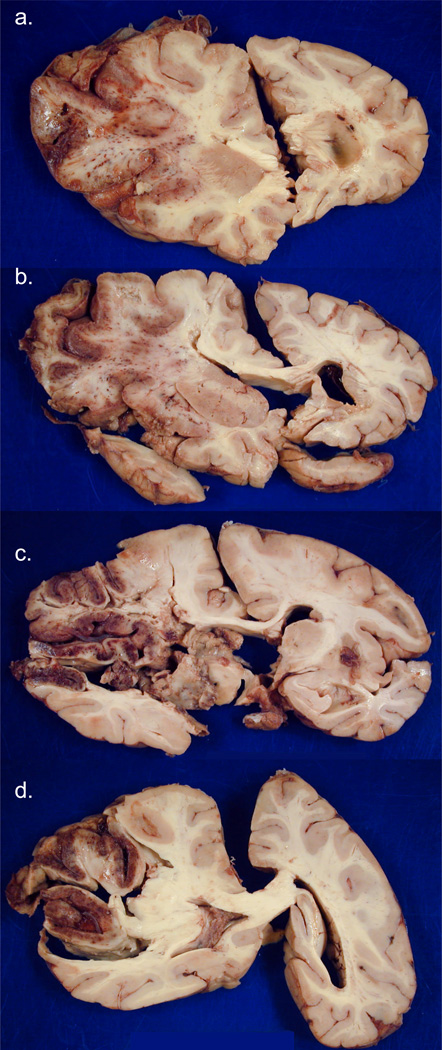
FIGURE 2.
Case #2. [Photographs of brain slices in this and subsequent figures follow the neuropathology convention of left side of the brain appearing on the left of the image.] Coronal sections of fixed slices from cerebral hemispheres arranged rostral to caudal in a-d show subacute necrosis and edema on the left, with significant left to right shift of midline structures. The infarct is extensively hemorrhagic and involves most of the left MCA territory..

