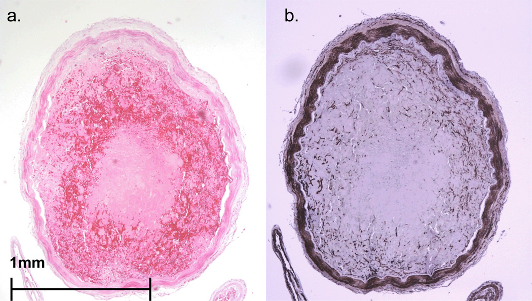
FIGURE 6.
Case #3. Representative cross section of right MCA (A from section stained with H&E, B from a parallel section immunostained with primary antibody to SMA) showing a well organized thromboembolus in a relatively non-atherosclerotic artery. Panel B highlights ingrowth of smooth muscle cells into the thromboembolus.

