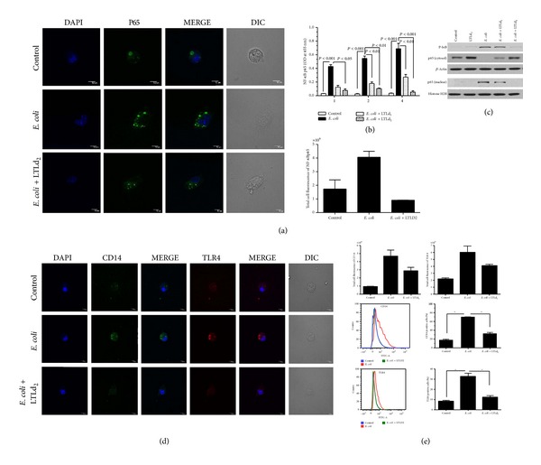
Figure 3.

Effect of LTL on expression levels of (a) NF-κB p65 in peritoneal macrophage cells after 4 h incubation at concentration LTLd2 (100 μg/mL) in the presence and absence of heat-killed E. coli (O18:K1; 1 × 10⁸ CFU/mL), from immunofluorescence level measured by confocal laser scanning microscopy. (b) ELISA results showed the nuclear extract of NF-κB p65 in peritoneal macrophage cells after 1, 2, and 4 h incubation with LTLd1 and LTLd2. (c) Western blot analysis revealed nuclear and cytosolic NF-κB p65 levels and p-IκB protein expression level after treatment with LTLd1 and LTLd2 following 12 h incubation. (d) Immunofluorescence expression measured by confocal laser scanning microscopy (magnification 600x) and (e) by flow cytometry showing levels of CD14 and TLR4 in peritoneal macrophage cells after 4 h incubation at concentration LTLd2 in the presence and absence of heat-killed E. coli (O18:K1; 1 × 10⁸ CFU/mL). The data are reported as the mean ± SEM of triplicate experiments (*P < 0.05, **P < 0.01).
