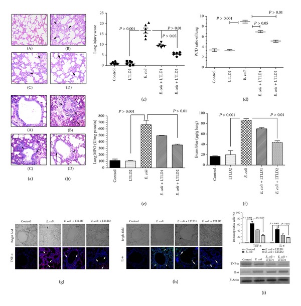
Figure 5.

Effects of LTL on lung histopathological changes in heat-killed E. coli challenged mice (n = 10). Mice were administered i.p. with LTL at doses LTLD1 (25 mg/kg) and LTLD2 (50 mg/kg) prior to heat-killed E. coli O18:K1, 104 CFU challenge. (A) Control group, (B) E. coli group, and (C) and (D) with LTL at doses LTLD1 and LTLD2. The arrows indicate infiltration generated prominent inflammatory cells and alveolar hemorrhage. Left panel (a) shows H&E staining and right panel (b) shows PAS staining, original magnification (×200). (c) The total lung injury score was calculated by adding up the individual scores of each category. (d) Lung W/D ratio assessment and the evaluation (e) of lung MPO activity with (f) the extravasation of parenchymal vascular leak. Localization of TNF-α (g) and IL-6 (h) in lung tissues after E. coli challenge in mice. Magnification (200x) arrows indicate immunopositive cells. Approximately 200 cells were counted per field, five fields were examined per slide, five slides were examined per group, and data were validated by western blot analysis (i). Values are presented as mean ± S.E.M. (*P < 0.05, **P < 0.01).
