
Figure 3.
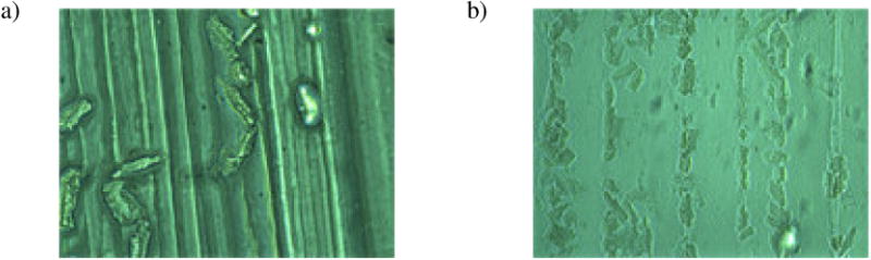
Phase-contrast microscopic images of feline adult cardiomyocytes on the printed alginate channels. Poor attachment was seen on pure alginate channels (a); on the contrary, many cardiomyocytes were found to align and attach to alginate/laminin channels (b).