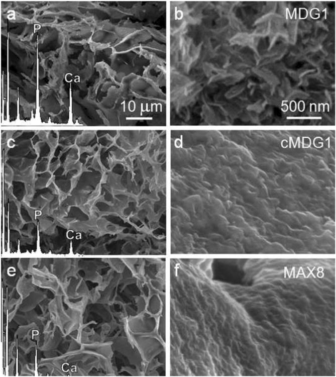
Figure 4.

SEM micrographs and corresponding EDS spectra of lyophilized MDG1 (a), cMDG1 (c), and MAX8 (e) gels after 24 hr mineralization. The morphological differences among the deposited minerals are observable at higher magnifications in panels b, d and f, respectively.
