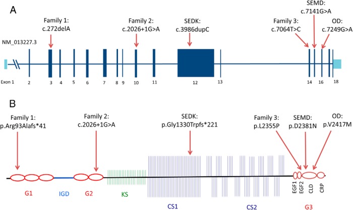
Figure 4.
Structure of the ACAN gene and aggrecan protein and the locations of current and previously reported mutations. A, Top panel shows the gene structure (RefSeq NM_013227.3) of ACAN and the locations of the variants identified in Families 1, 2, and 3, as well as the previously identified pathogenic variants (SEDK, spondyloepiphyseal dysplasia type Kimberly; SEMD, spondyloepimetaphyseal dysplasia; OD, osteochondritis Dissecans); B) Organization of the aggrecan proteoglycan (G1, 2, 3, globular domain 1, 2, 3; IGD, interglobular domain; KS, keratan sulfate; CS1, 2, chondroitin sulfate 1, 2; CLD, C-type lectin domain; CRP, complement regulatory like domain; EGF1, 2, epidermal growth factor–like domain 1, 2). Protein coordinates are based on RefSeq NP_037359.3. Figures are modified from (29) and are drawn approximately to scale.

