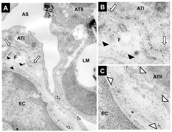
Figure 4.
An electron micrograph (EM) of normal lung tissue showing microdomains in the extracellular matrix which contain varying amounts of sulfation. Cytochemical visualization of sulfation in the extracellular matrix was performed using high iron diamine (HID), which binds to sulfates in lung tissue. The HID reaction product forms discrete 5–12 nm silver particles following appropriate intensification and is seen as discrete dark spots (Sannes, 1984). The basement membrane under a type I cell and an adjacent fibroblast (F) is highly sulfated (Arrows in 2A and 2B). The microdomains surrounding the pulmonary fibroblast show this cell to have microdomains of high and low sulfation. The extracellular matrix adjacent to the ATI cell is highly sulfated (white arrows in 2A and 2B) whereas the extracellular matrix adjacent to the endothelial cell (EC) is undersulfated (black arrowheads in 2A and 2B). In contrast to the ATI cell, this EM shows that the extracellular matrix underlying the type II cell (ATII) is undersulfated. White arrowheads in 2A and 2C define the extracellular matrix underlying the ATII cell and a pulmonary capillary EC (This figure is from studies by Frevert, et al., (Frevert and Sannes, 2006) with permission.).

