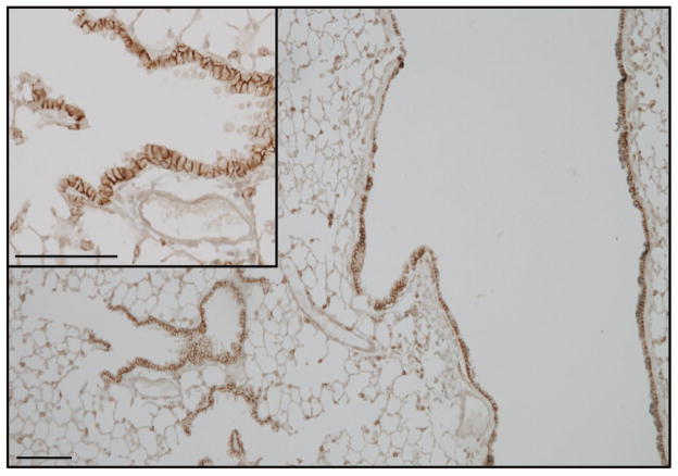
Figure 5.
Positive staining (brown) for syndecan-1 (syndecan-1) in normal mouse lungs shows a basal lateral distribution in airway epithelial cells and immunoreactivity in cells of the alveolar septa. Immunohistochemistry for syndecan-1 was performed with a rat anti-mouse syndecan-1 IgG (BD/Pharmingen, Franklin Lakes, NJ).

