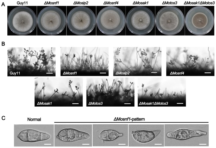
Figure 2. Comparison of the SNF1 pathway mutants with regard to colony morphology and conidial development.
(A) Strains were cultured on CM plates at 25°C for 10 days. ΔMosak1ΔMotos3 exhibited a decreased mycelial growth rate, while no significant difference in the colony size was observed between other mutants and Guy11. (B) Microscopic observation of conidial development. Significant reduction in conidial production was observed in ΔMosnf1, ΔMosip2, ΔMosnf4, ΔMosak1, and ΔMosak1ΔMoto3 at 24 hpi. However, ΔMotos3 developed short, yet dense conidiophores with plenty of spores arrayed thereon. Bars = 50 µm. (C) Conidia of WT and the mutants were harvested and observed under the light microscope. Conidial shape of ΔMosip2, ΔMosnf4, ΔMosak1, and ΔMosak1ΔMoto3 was identical to that of ΔMosnf1 (ΔMosnf1-pattern), whereas there was no measurable difference between ΔMotos3 and Guy11 (Normal). Bars = 5 µm.

