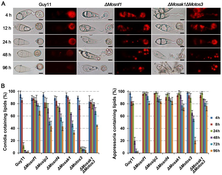
Figure 6. Intracellular mobilization of lipid droplets in WT and SNF1 pathway mutants during appressorium morphogenesis.
Conidial suspensions were incubated on the surfaces of hydrophobic films and stained with Nile red to observe the status of lipid droplets movement and distribution at the indicated time points under epifluorescence microscope. (A) ΔMosnf1 and ΔMosak1ΔMotos3 showed significant delays in lipid mobilization and degradation with the presence of Nile red-stained lipid bodies even at 96 hpi, while fluorescent signals were almost invisible in WT at 48 hpi. Bars = 5 µm. (B) Percentages of conidia (left) or appressoria (right) that contained lipid droplets. Varied degrees of defect in lipid mobilization were observed among the mutants.

