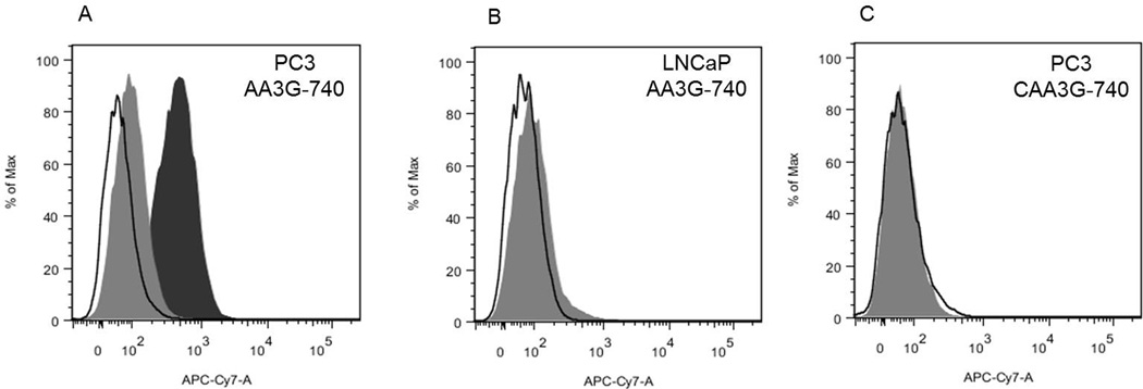
Figure 2. Binding specificity.
A) AA3G-740 efficiently labeled PC3 cells that have high level of GRPR (black filled area). Co-incubation with bombesin prevented the binding, resulting in minimal cell fluorescence (gray filled area). B) Low GRPR expressing cells, LNCaP, showed minimal increase in fluorescence (grey filled area). C) Control agent, CAA3G-740 containing a scrambled binding sequence was not able to label PC3 cells (grey filled area). Black outline represent unstained cells in all three panels.

