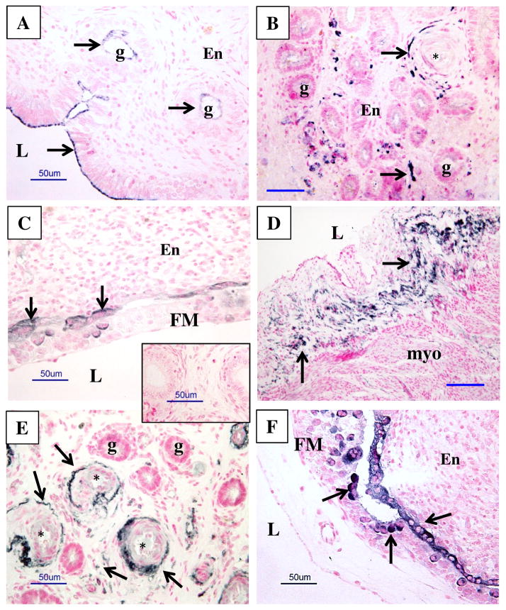
Figure 6.
Representative images of PrPC protein localization (dark staining) in uterine and uteroplacental tissues of nonpregnant (NP; A and B) ewes and pregnant ewes from days 20–30 after mating (C–F). Note a lack of positive staining in control (inset in C), where primary antibody was replaced with mouse IgG. Arrows indicate some areas of PrPC-positive (dark) staining; the counterstaining (pink) was with nuclear fast red. L = lumen of uterus, g = uterine gland, En = endometrium, FM = fetal membranes (chorioallantois), myo = myometrium, * = blood vessels. Note the presence of PrPC protein in NP at the apical borders of the luminal epithelia and epithelia of luminal glands, larger blood vessels (primarily vascular smooth muscle and adventitia) and in some cells in stromal tissues of endometrium (A and B), and in tissues from pregnancy in luminal epithelia adjacent to the FM, in luminal epithelial cells of maternal caruncle (F; arrow), in bi- and multinucleated cells of the placenta (F; arrowheads), and in larger blood vessels and some cells in stromal tissues (E). Size bars = 50 μm.

