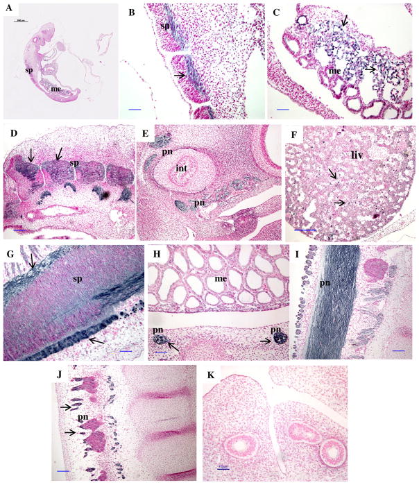
Figure 7.
Representative images of immunolocalization of PrPC protein in a longitudinal section of an embryo on day 20 (A), and fetal tissues from days 20 (B, C), 22 (D), 24 (E), 26 (F), 28 (G) and 30 (H–J) of early pregnancy. Note a lack of positive staining in control (K) where primary antibody was replaced with mouse IgG. Arrows indicate areas of PrPC positive (dark) staining in contrast to the background (pink) stained with nuclear fast red. sp = spinal cord, pn = peripheral nerves, liv = liver, int = intestine, me = mesonephros. Note the presence of PrPC in the spinal cord (B, D, G), peripheral nerves associated with the intestine and other peripheral nerves (E, H, J, I), liver (F), and mesonephros (C). Size bars = 500 μm on A, 100 μm on B–E and G–K, and 200 μm on F.

