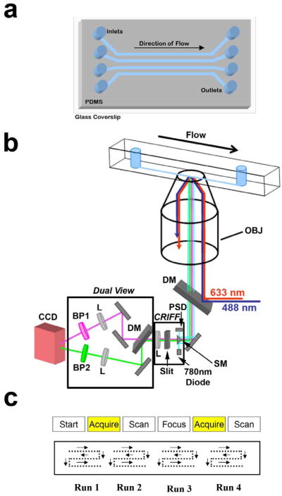
Figure 3. Imaging isolated cellular compartments.
(a) We use a microfluidic chip fabricated in PDMS and sealed to a glass coverslip with four large (800μm (w) × 200μm (h) × 2 cm (l)) channels to image samples. (b) Schematic showing the optical path for the TIRF microscope coupled with the CRIFF and dual view. Abbreviations: Lens (L), Dichroic Mirror (DM), Continuous Reflective-Interface Feedback Focus System (CFIFF), Charged Coupled Device (CCD), Position Sensitive Diode (PSD), Scanning Mirror (SM), Band Pass Filter (BP). (c) Diagram showing the scanning and acquisition pattern we used to collect images along the channel.

