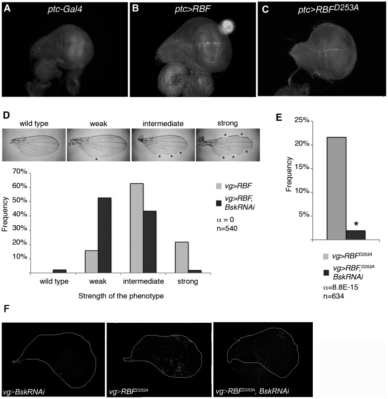
Figure 5. RBF- and RBFD253A-induced apoptosis as well as RBFD253A-induced overgrowth depend on the JNK pathway activity.
(A–C) Anti-◯P-JNK staining in young third instar larvae wing imaginal discs. (A) No staining is observed in ptc-Gal4/+ control. (B, C) UAS-RBF/ptc-Gal4 and UAS-RBFD253A/X; ptc-Gal4/+ discs present anti-◯P-JNK staining at the dorso-ventral boundary. All discs are shown with posterior to the top. (D) Notch phenotypes in adult wings of vg-Gal4/+; UAS-RBF and vg-Gal4/+; UAS-RBF/UAS-bsk-RNAi flies are grouped into four categories (wild type, weak, intermediate and strong) according to the number and size of notches observed on the wing margin (asterisks). Bsk-RNAi partially suppresses RBF-induced notch phenotypes (Wilcoxon test, α<10−15, n = 540). (E) The frequency of RBFD253A-induced ectopic tissue growth is strongly decreased in UAS-bsk-RNAi co-expressing flies (Chi2 test, α = 8.8 10−15). (F) TUNEL-labeling of apoptotic cells in wing imaginal discs; specific staining of apoptotic cells corresponds to bright patches in wing discs of vg-Gal4/+; UAS-bsk-RNAi/+ (left panel), vg-Gal4/+; UAS-RBFD253A/+ (center panel), vg-Gal4/+; UAS-RBFD253A/UAS-bsk-RNAi (right panel) larvae. RNAi-mediated knockdown of bsk strongly decreased RBFD253A-induced apoptosis. All discs are shown with posterior to the top.

