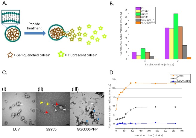
Figure 5. The induce of calcein leakage and liposome disruption with amyloidogenic peptides.
(A) Upon the disruption of the membrane, the self-quenched calcein is released from the liposome and generate strong fluorescence signal. (B) The induced calcein leakage (%) was monitored in the presence of different peptides after 30 or 60 minutes incubation. (C) Freshly extruded large unilamellar vesicles (LUV) were identified either alone (left) or in the presence of G295S (middle) or GGG308PPP (right). The scale bars represent 100 nm. (D) Time-course of calcein fluorescence enhancement in the presence of D1, G295S, and GGG308PPP (50 µM).

