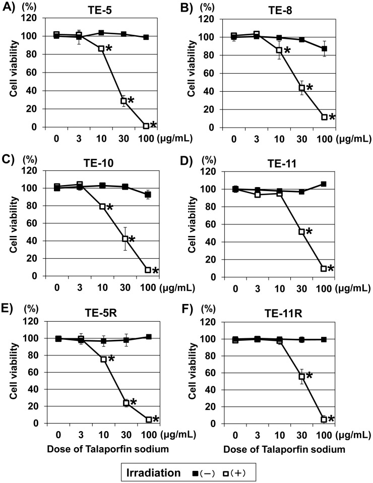
Figure 2. Cytotoxic effect of t-PDT on ESCC cells.
Cell viability at 48-PDT was assessed using the WST-1 assay. t-PDT induced a talaporfin sodium dose-dependent cell death in ESCC cells (white square in the figures) regardless of differentiation grade or 5-FU resistance. (A) TE-5 (derived from poorly differentiated ESCC), (B) TE-8 (derived from moderately differentiated ESCC), (C) TE-10 (derived from highly differentiated ESCC), (D) TE-11 (derived from moderately differentiated ESCC), (E) TE-5R (5-FU-resistant cells derived from parental TE-5 cells) and (F) TE-11R (5-FU-resistant cells derived from TE-11 cells). A viability of 100% was defined as the amount of absorption at 450 nm found in untreated (non-irradiated and absence of treatment with talaporfin sodium) cells. Each point represents the mean ± S.D. from experiments conducted at least in triplicate. *P<0.01 vs untreated (non-irradiated and absence of treatment with talaporfin sodium) cells (n = 3).

