
Figure 2.
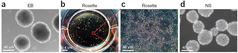
Step-by-step illustration of neuroepithelial differentiation. (a) Under a chemically defined system, hPSCs form EBs in suspension culture for the first 7 d. (b) The EBs are then plated on day 7 at the density as shown. (c) By day 10, each EB will develop into a colony that contains neuroepithelial cells in the form of rosettes. (d) At day 16, the rosette-containing colonies are detached and grown in suspension to form neuroepithelial spheres (NS).