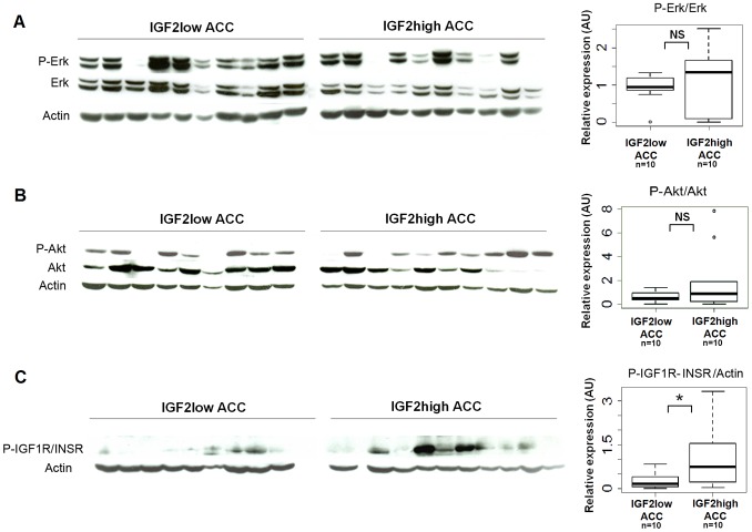
Figure 3. IGF signaling pathway activation in IGF2-high and IGF2-low ACC.
The activation of Erk (A), Akt (B), and IGF1R/INSR receptors (C) was analyzed by western blotting with antibodies directed against the phosphorylated form of these proteins, in IGF2-high (n = 10) and IGF2-low (n = 10) ACC. Boxplots show the quantification of the results of western blots. Y-axis: results of the quantification of the western blot bands normalized to total Erk, Akt or actin. Wilcoxon test results (*p<0.05; NS = not significant) are indicated. The phosphorylation of the receptors is higher in IGF2-high than in IGF2-low ACC, whereas the activation of Erk and Akt downstream pathways is similar.

