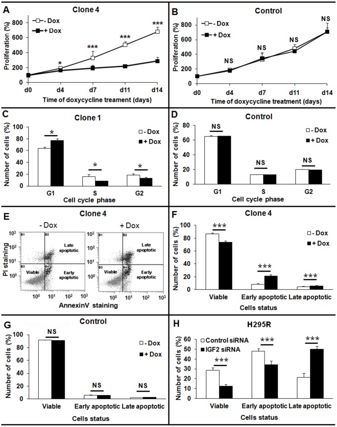
Figure 4. Consequences of IGF2 knock-down on cellular growth, apoptosis, and the cell cycle in H295R cells.
A, B: Consequences of long-term knock-down of IGF2 on H295R cell growth as assessed by a MTT proliferation test. A: Doxycycline treatment leads to IGF2 knock-down and to a significant impairment of cellular proliferation in a clone with integrated IGF2 shRNA. Results are presented for clone 4, which showed the most significant impairment of cell growth B: Doxycycline (black square) does not inhibit proliferation in a control clone. C, D: Effect of IGF2 knock-down on the cell cycle. After 7 days of doxycycline treatment, the cell cycle was analyzed by PI staining and flow cytometry. Cells expressing IGF2 shRNA were largely arrested in the G1 phase of the cell-cycle and few cells were in S phase (C). Results are presented for clone 1, which showed the most significant G1 cell cycle arrest. Doxycycline treatment did not affect the cell cycle of a control clone (D). E-H: Apoptosis studied by flow cytometry after PI (Propidium iodide) and Annexin V staining. E: Example of FACS results for clone 4, showing the apoptotic status of cells according to Annexin V (X axis) and PI (Y axis) staining. F, G: Study of non-induced apoptosis after prolonged IGF2 knock-down. Ten days after shRNA induction by doxycycline treatment, cells were stained and analyzed by flow cytometry. Doxycycline treatment did not affect apoptosis of the control clone without integrated shRNA (G). Both early and late apoptosis were significantly higher in cells expressing IGF2 shRNA than in control cells (F). Results are presented for clone 4, which showed the most significant difference in apoptosis. H: Study of TNF-alpha-induced apoptosis after the transient knock-down of IGF2 in H295R cells. Cells were treated with TNF-alpha 48 h after siRNA transfection, and were analyzed by flow cytometry 48 h later. The number of viable cells was significantly lower in cells transfected with siRNA against IGF2 (black bars), and the percentage of cells in late apoptosis was significantly higher than in cells transfected with a control siRNA (white bars). Results were analyzed with the Wilcoxon test. *: p<0.05. ***: p<0.001. All the results presented in this figure are representative of at least three independent experiments.

