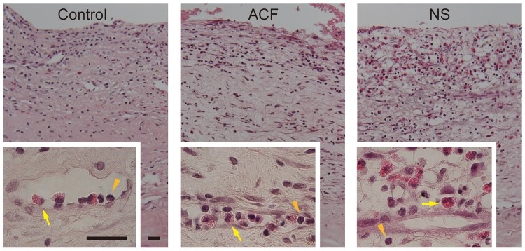
Figure 2. Cross sections of the hematoma outer membranes fixed immediately after surgery (Control, left panels) or after 24 h of incubation with artificial cerebrospinal fluid (ACF, center panels) or normal saline (NS, right panels).
The insets are higher magnifications of the perivascular area. Inflammatory cells, including lymphocytes (orange arrowheads) and eosinophils (yellow arrows), are mostly located in the loose stroma of the hematoma membrane in the NS group, whereas they are confined in the capillary lumen, in the control and ACF groups. Hematoxylin–eosin stain, Burr = 25 µm.

