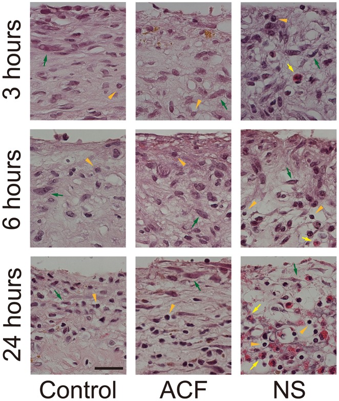
Figure 3. The membranes were fixed after 3, 6, or 24 h of incubation.
Fewer morphological changes are seen in the control (left panels) or ACF groups (center panels), whereas the cytoplasm of constituent cells shrank and the nuclei of most infiltrated cells are pyknotic and deformed (round) in the NS group (right panels). The former alteration is the most noticeable in fibroblasts (green arrows) and the latter in lymphocytes (arrowheads).

