
Figure 7. Protein expression of CASK, NRF2 and SNCA in rat hippocampus after TBI.
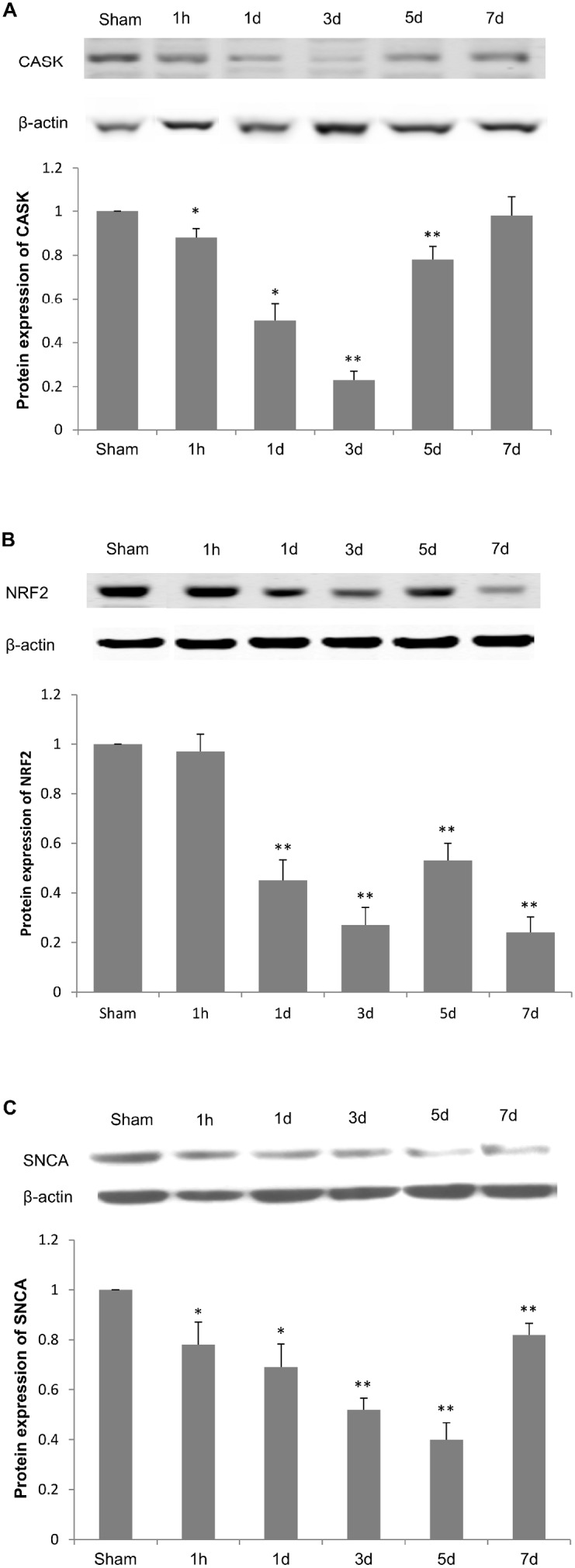
Western blots were used to detect CASK (A), NRF2 (B) and SNCA (C) protein levels in whole cell lysates of hippocampus. Relative protein expression levels were determined by normalizing band density of target protein to that of β-Actin and comparing with sham group. Data are presented as mean ± SEM. N = 5. *P<0.05 and **P<0.01 vs. sham group.