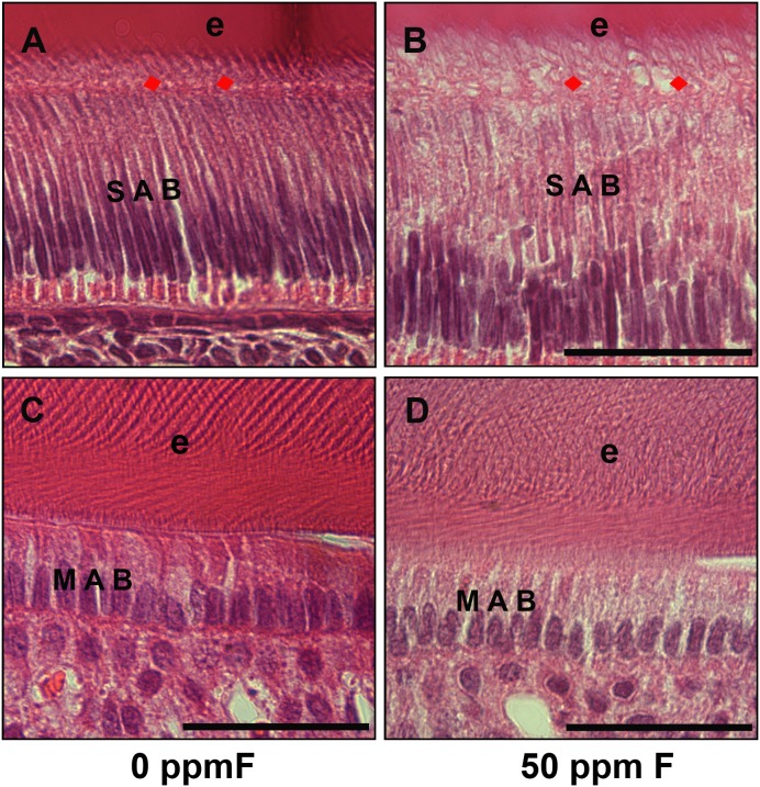
Figure 2. H&E stained histological sections from control and fluoride treated mouse incisors.
A) H&E stained control mouse incisors showed elongated and polarized secretory ameloblasts (SAB) and clearly aligned Tomes’ processes (TP) (red arrows). B) Secretory ameloblasts (SAB) in the fluorosed incisors were less organized, had increased amounts of clear vacuoles in cytoplasm. Tomes’ processes (TP) (red arrows) were less distinct as compared to controls. The space between ameloblasts and enamel layer was filled with increased numbers of vacuoles and was obviously wider in the fluorosed incisors. C) H&E stained mouse control maturation ameloblasts (MAB) and mineralizing enamel (e). D) Morphology of the maturation amelobasts (MAB) in the fluorosed mouse incisors showed no obvious differences as compared to controls. Scale bar: 100 µm.

