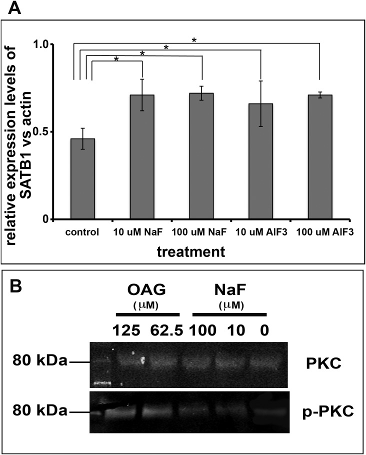
Figure 6. Ameloblast-lineage cells had increased p-PKC and SATB1 when they were exposed to fluoride.
A) Densitometry analysis of protein extracted from human ameloblast-lineage cells grown in media supplemented with NaF or AlF3 shows significant increase in SABT1 protein when cells were exposed to either 10 or 100 µM fluoride as compared to control. One way ANOVA shows P<0.05, Tukey’s multiple comparison post-test shows * is significantly different from the control samples (n = 4). B) The Western blot analysis showed that OAG, the analog of DAG, at concentration of 62.5 µM or 125 µM significantly increased the phosphorylated PKC, which served as a positive control. NaF at 10 µM or 100 µM also increased the levels of phosphorylated PKC in ameloblast-lineage cells, though at the moderate levels as compared to OAG treated cells. Either OAG or NaF did affect the levels of PKC.

