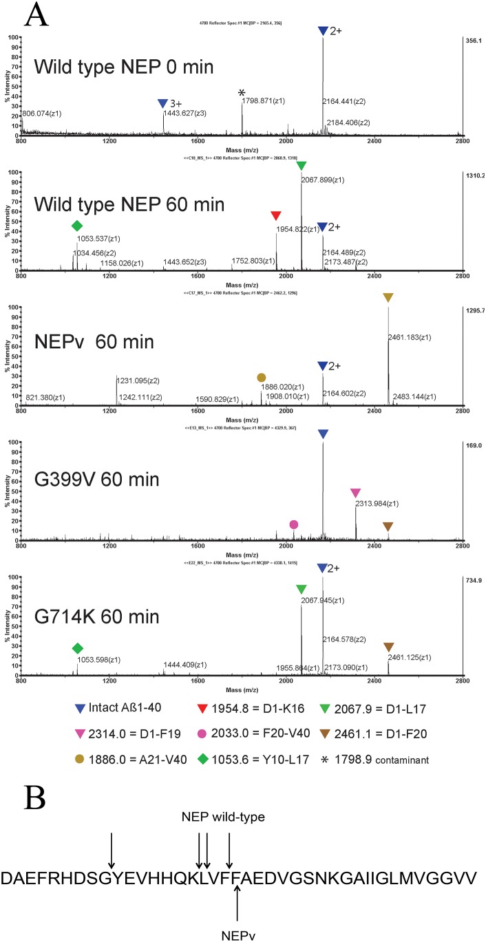
Figure 4. MS analysis of Aβ1–40 peptide cleaved by wild type NEP and variants.
Aβ1–40 peptide was incubated with different sequence variants of neprilysin and the appearance of cleaved fragments was detected by MALDI mass spectrometry. A) expanded region between 800–2800 m/z. Peptide identities were confirmed by MS/MS analysis and are indicated by triangles (N-terminal fragments) or circles (C-terminal fragments). B) Cleavage preference on Aβ1–40 exhibited by wild type NEP and NEPv. Arrows indicate the predominant sites of cleavage observed.

