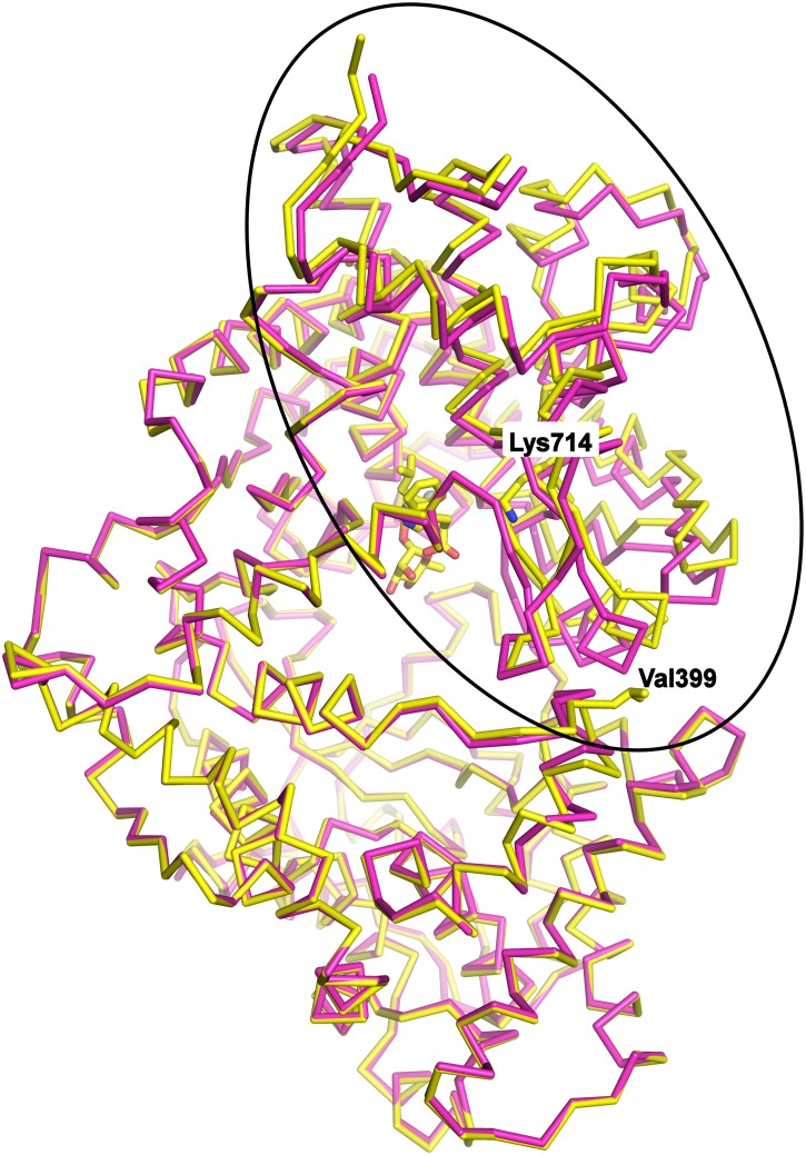
Figure 5. Conformational change of NEPv.
Superposition of NEPv (yellow) with the published wild-type NEP structure 1dmt (magenta) (reference). The main effects of the G399V/G714K mutations are observed within the encircled part of the protein. The upper half is domain 1, the lower half is domain 2, and linker segments connecting the domains are to the left. The positions of Val399 and Lys714 are indicated and the mutants and phosphoramidon of NEPv are shown in sticks. The zinc ion is shown as a grey sphere. All pictures and structure alignments were made using Pymol.

