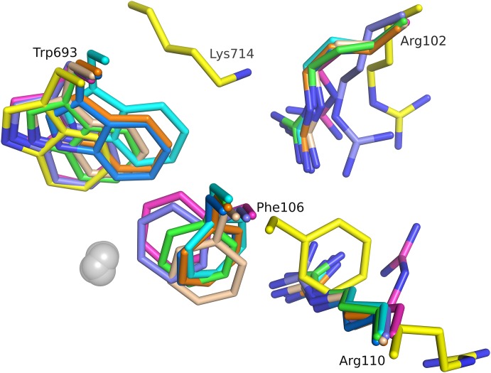
Figure 6. Conformational changes of residues involved in substrate binding.
Superpositions of NEPv (yellow) with published structures 1 dmt (magenta), 1r1h (cyan), 2yb9 (violet), 1r1i (green), 1r1j (wheat), 1y8j (blue) and 2qpj (orange). Large ligand induced flexibility in active site. Flexible side chains include Arg102, Phe106, Arg110 and Trp693. Ligands left out for clarity. Zinc ions shown as grey spheres.

