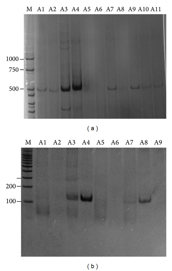
Figure 2.

Dengue virus amplification by RT-PCR from blood samples patients. (a) Electrophoretic profile of DNA fragments (511 pb) corresponding to positive samples. A5, A6, A8: negative samples for Dengue virus. (b) Electrophoretic profile from the nested PCR for viral typing. A3, A4, A7, A8: DENV-2 fragment of 119 pb. PCR products were fractioned by 8% PAGE and visualised by silver staining. M: molecular size markers (bp).
