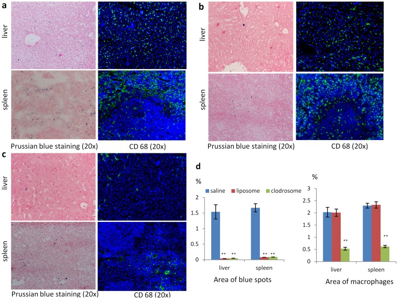
Figure 5. Prussian blue staining and CD68 immunostaing of liver and spleen in saline (a), liposome (b) and clodrosome (c) group.
The blue dots on Prussian blue staining are the liver-accumulated SPIO nanoparticles; the green fluorescence on fluorescence images is macrophage. d. The area occupied by kupffer cells and SPIO nanoparticles estimated by imaging analysis in liver and spleen. (**p<0.005 compared with saline group. n = 4).

