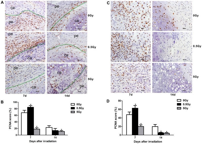
Figure 5. Cell proliferation during intramembranous osteogenesis and chondrogenesis.
PCNA immunostaining was performed at 7 and 14 days after X-ray irradiation. Low-dose X-ray irradiation promoted the proliferation of periosteum cells and chondrocytes at day 7 after irradiation in fracture sites, whereas high-dose X-ray irradiation inhibited the proliferation of these cells at both days 7 and 14 post-irradiation (A, C). Quantification of cell proliferation in each group (B, D). The green dot represents the demarcation between the soft tissue and cartilage, and the white arrow denotes a vessel. pe: periosteum, ca: cartilage, n = 4 rats/group,*p<0.05 vs. the control group.

