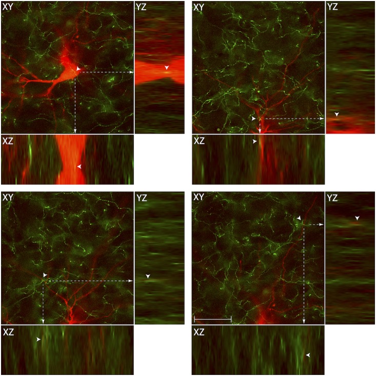
Figure 5. Close apposition between chemically-identified axons and thalamic trigeminovascular neurons.
Images from the original z-stack (obtained every 1 µm) were used to create orthogonal views in the y–z and x–z planes. The three views provide evidence that SERT immunopositive fibers (green) may contact cell bodies, proximal and distal dendrites of trigeminovascular neurons in Po (red; as shown in Fig. 4). Note that some green-labeled axons and red-labeled soma or dendrites are in the same focal plane (yellow). To see similar images for all the neurotransmitters and neuropeptides identified in this study, see Supplementary Figures 1–7. Caveat: proximity between the chemically-identified axons and the TMR-labeled trigeminovascular thalamic neurons suggests that they are innervated by the different neuropeptides/neurotransmitters. Definitive evidence for actual synapses, however, requires tissue examination with electron microscopy. Scale bar = 50 µm.

